Standard breast cancer checks are missing the full picture for millions of patients, leaving conventional screening protocols dangerously outdated in the face of biological reality. Traditional mammography remains the undisputed anchor of public health policy. Yet, looking closely at population-wide data reveals a glaring discrepancy between standard screening guidelines and individual anatomical safety. Millions of women dutifully attend their annual or biennial appointments, receive a clean bill of health, and trust the machinery. Months later, a palpable mass emerges. The system calls it an interval cancer. Clinicians whisper about limitations. Patients are left to pick up the pieces.
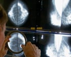
Current screening protocols are built on broad statistical averages rather than personal physiological risk. This is the structural flaw hiding in plain sight. Mammograms work by passing X-rays through compressed breast tissue to capture grayscale images. Dense tissue, however, appears white on a mammogram. Cancerous tumors also appear white on a mammogram. Hiding a tumor inside dense tissue is mathematically equivalent to hiding a snowball in a blizzard.
Approximately forty percent of women have dense breasts. For this massive segment of the population, standard mammography loses a significant portion of its diagnostic sensitivity. We are relying on a tool that struggles to see through the very tissue it is examining.
The Density Dilemma and the Limits of 2D Imaging
To understand why traditional checks fall short, we must examine the physics of the equipment. Standard two-dimensional mammography flattens three-dimensional structures into a single flat image. Overlapping tissue layers obscure abnormalities. A slow-growing lesion can easily hide behind a benign fibrous ridge for years.
Digital breast tomosynthesis, often called three-dimensional mammography, represents an upgrade by taking multiple low-dose X-rays from different angles. This slices the breast into millimeter-thick layers, reducing the camouflage effect of overlapping tissue. Yet, tomosynthesis is not a universal cure. While it improves cancer detection rates and reduces false positives, it still relies on X-rays. It still struggles when tissue density is extremely high.
Radiation exposure also remains a background concern, albeit a low one relative to the benefits of early detection. More importantly, the psychological toll of false positives cannot be overstated. A callback for an additional view triggers weeks of acute anxiety, unnecessary biopsies, and financial strain, all because a shadow on a grainy image looked suspicious.
Patients deserve better than a guessing game dictated by tissue density.
The High Cost of One-Size-Fits-All Guidelines
Public health organizations issue broad guidelines for a reason. Population-level mandates are cheap to administer, easy to communicate, and straightforward to track. Starting mammograms at age forty or fifty works well on a spreadsheet. In human practice, biology refuses to read the rulebook.
Aggressive, fast-moving tumors do not wait politely for a woman to turn forty. Triple-negative breast cancers and other high-grade malignancies frequently strike younger patients, growing rapidly in the interval between scheduled two-year screenings. By the time a routine check rolls around, the cancer has advanced to stage three.
Meanwhile, low-risk patients undergo decades of routine imaging that might catch indolent ductal carcinoma in situ that would never have threatened their lives. This leads to overtreatment, surgery, and radiation for conditions that might have remained harmless.
We have optimized our screening infrastructure for statistical efficiency rather than diagnostic precision.
Integrating Ultrasound and MRI Into the Front Lines
If standard mammography hits a wall with dense tissue, why is supplemental screening still treated as an elite luxury rather than a standard option?
Automated and handheld breast ultrasound technologies offer a vital window for women with dense breasts. Ultrasound uses sound waves rather than ionizing radiation. It can spot solid masses hiding inside dense tissue that completely elude an X-ray. The catch is a higher false-positive rate. Ultrasound operators require specialized training, and the equipment is less standardized than mammography units.
Magnetic resonance imaging provides the gold standard for high-risk surveillance. Contrast-enhanced MRI visualizes blood flow to rapidly dividing cells, making it extraordinarily sensitive for detecting hidden malignancies.
Consider a hypothetical patient named Sarah. Sarah has extremely dense breast tissue and a family history of the disease, but carries no known genetic mutation like BRCA1 or BRCA2. Under standard insurance guidelines in many jurisdictions, her dense tissue alone does not qualify her for a screening MRI. She gets a standard mammogram, gets told everything is clear, and goes home. Her risk profile remains invisible to the algorithm.
If Sarah had access to automated breast ultrasound as a mandatory adjunct, her outcome might change entirely.
Overhauling the Risk Assessment Model
Fixing this broken system requires abandoning the calendar-based approach to screening. Age alone is a lazy proxy for risk.
Modern medicine possesses the tools to stratify patients accurately before they ever step into a clinic. Polygenic risk scores analyze millions of single nucleotide polymorphisms across the genome to calculate an individual's lifetime susceptibility to breast cancer. Combined with lifestyle factors, hormonal history, and precise volumetric breast density measurements, clinicians can build a dynamic risk profile.
A twenty-eight-year-old with a high polygenic risk score and dense tissue should be flagged for early, intensive surveillance involving alternating MRIs and mammograms. A sixty-year-old with low density and low risk might safely transition to less frequent checks.
Insurance reimbursement models must shift to match this reality. Payers currently balk at covering supplemental ultrasound or MRI for average-risk women with dense breasts, citing cost. They ignore the downstream financial and human cost of treating advanced, metastatic cancer that could have been caught years earlier at stage zero or one.
The conversation around breast cancer checks must move past the comforting illusion that a biennial mammogram is a universal shield. It is a blunt instrument. Acknowledging its limits is the only path toward building a screening architecture that actually protects every single patient.