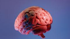
Western science doesn't understand your brain. That sounds like hyperbole, but it's the stark reality facing global neuroscience right now. For decades, the standard atlas used by neurosurgeons and researchers worldwide has been based on Caucasian brains. We assumed a brain is a brain, regardless of ethnicity. We were wrong.
Indian brains are structurally different. They are smaller in height, width, and volume compared to Western counterparts. If a surgeon uses a Western template to operate on an Indian patient, the margin of error increases. This isn't just an academic problem. It affects misdiagnosis rates for conditions like Alzheimer's and changes how we plan delicate brain surgeries.
That's why neuroscientists in India stopped waiting for the West to catch up. They started building their own maps.
The Indian Brain Atlas Project Breaks the Mold
The International Consortium for Brain Mapping (ICBM) template has long been the gold standard. But this standard relies heavily on data from Caucasian subjects. When researchers at the International Institute of Information Technology, Hyderabad (IIIT-H) analyzed MRI scans of Indian subjects, the discrepancies were glaring. The MNI152 template, a widely used standard from the Montreal Neurological Institute, simply didn't fit.
Led by Jayanthi Sivaswamy, the IIIT-H team created the first population-specific Indian brain atlas, known as IBA100. They validated what many suspected. The Indian brain is significantly smaller in three dimensions. The structural differences mean that using a Western atlas can lead to a misallocation of brain regions during automated MRI analysis.
Think about what happens during an early-stage Alzheimer's scan. Doctors look for the shrinking of the hippocampus. If the software uses a Caucasian baseline, it might flag a perfectly healthy, naturally smaller Indian hippocampus as atrophied. That leads to false positives, unnecessary panic, and wrong treatment plans.
Moving From Structural Coordinates to Cellular Architecture
Mapping a brain isn't just about measuring its outer boundaries. It requires understanding the dense networks inside. The National Brain Research Centre (NBRC) in Manesar took the mapping mission further by looking at the neuroanatomy of the aging Indian population.
Their work focuses on identifying early biomarkers for neurodegenerative diseases. By establishing a baseline of how a healthy Indian brain ages, they can spot the exact moment things start going sideways. The project relies on structural MRI, functional MRI (fMRI), and magnetic resonance spectroscopy (MRS).
MRS is particularly useful here. It lets researchers look at the chemical composition of brain tissue without cutting anything open. The NBRC team tracks glutathione, a critical antioxidant in the brain that drops significantly in Alzheimer's patients. By monitoring this decline against an Indian-specific norm, they catch cognitive decline years before external symptoms show up.
The Technological Hurdle of Processing Petabytes of Data
The human brain holds roughly 86 billion neurons. Mapping the connections between them creates an astronomical amount of data. You can't process this on a standard laboratory computer.
Indian institutes had to upgrade their computational infrastructure to handle the load. The Indian Institute of Science (IISc) in Bengaluru deployed high-performance computing clusters to manage high-resolution diffusion tensor imaging (DTI) datasets. DTI tracks the movement of water molecules along white matter tracts, revealing the brain's underlying wiring.
The sheer scale of data processing is where many neuroimaging projects stall out. A single high-resolution brain scan can take up gigabytes of space. Multiply that by thousands of subjects across different age groups and demographics, and you are dealing with petabytes of data. The IISc teams rely on specialized algorithms to clean up the noise inherent in MRI scans, ensuring the final maps are accurate enough for clinical use.
Why Localized Neuroscience Saves Lives
This isn't just about national pride. It's about precision medicine. The variance in brain structure affects how brain tumors are resected.
When a neurosurgeon operates, they need to know exactly where the functional areas—like speech and motor control— reside. A shift of just a few millimeters can mean the difference between removing a tumor safely and leaving a patient unable to speak. Localized atlases give surgeons the exact coordinates they need for navigate the specific anatomy of the population they treat.
Furthermore, drug efficacy often links back to genetic and structural variations across populations. The way neurological disorders manifest in South Asia differs from the West due to lifestyle, genetics, and environmental factors. Having a localized baseline allows pharmaceutical researchers to design clinical trials that actually reflect the population using the drugs.
The Next Step for Medical Professionals and Researchers
If you are a clinician or researcher working in neurology, relying solely on legacy Western datasets is no longer defensible. The data is available, and the shift toward population-specific diagnostics is already happening.
Start integrating the IBA100 and subsequent NBRC datasets into your neuroimaging workflows. Update your analysis software to utilize these localized templates when processing scans for Indian patients. When evaluating cognitive decline or planning surgical interventions, validate your findings against the native structural baselines rather than defaulting to the standard MNI templates. The accuracy of your diagnoses depends on using the correct map.